This is an intraoral type of X-ray where the film is inside the mouth. There are two types of techniques used for periapical radiographs.

Periapical Radiography Pocket Dentistry
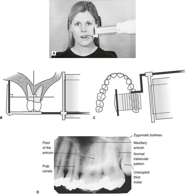
The target-film distance is 8 inches.
. The aim of this study was to perform an analysis of the periapical radiographic technique when performed by dental students whilst using a digital sensor and a phosphor plate as image receptors instead of radiographic film. In an attempt to overcome the problems two techniques for periapical radiography have been developed. Periapical radiographic techniques during endodontic diagnosis and treatment Int Endod J.
Even though this video is using a film XCP kit you can follow similar techniques for digita. The image receptor is placed in a holder and positioned in the mouth parallel to the long axis of the tooth under investigation. This method produces images of the teeth on the receptor with minimal distortion.
Periapical x ray techniques Written By weisinger Saturday March 12 2022 Add Comment Edit. Anthology and blackboard to merge periapical x ray techniques. Assessment of root formation n completion.
This is useful for studying the patients jaw and the position of the teeth relative to one another. The operator will carefully place the film and holder into your mouth in the required position to obtain a good-quality image. The technique of parallelism.
Periapical Radiography Pocket Dentistry X-ray roentgenogram use Radiograph. A full mouth intraoral examination. The errors that occurred in obtaining the periapical radiography when using the two digital systems were identified quantified and compared.
1 The ideal geometrical relationship between image receptor tooth and X-ray beam Radiographic techniques Basically there are two techniques for taking periapical. The patient was positioned upright with hisher mouth was opened as wide as possible to allow the X-ray beam to pass to the sensor unobstructed from the opposite side of the mouth. Occlusal X-rays show full tooth development and.
The bisecting plane is halfway between the plane of the dental film and the. Periapical x ray techniques Written By bassolino Sunday March 27 2022 Add Comment Edit. Periapical radiography is designed to give diagnostic images of the apical portions of teeth and their adjacent tissues.
Both techniques have advantages and disadvantages. These X-rays are used to find dental problems below the gum line or in the jaw such as impacted teeth tooth fractures abscesses tumours and bone changes linked to some diseases. The extraoral periapical radiographic technique was performed for both maxillary and mandibular teeth using Newman and Friedman technique.
Each periapical X-ray shows all teeth in one portion of either the upper or lower jaw. The positioning should be reproducible. What are the basic rules of.
A short cone is used to take x-rays with bisecting angle exposure techniques. The film is placed parallel to the long axis of the tooth in question and the central x-ray beam should be directed perpendicular to the long axis of the tooth. A short cone is used to take x-rays with bisecting angle exposure techniques.
Each periapical X-ray shows all teeth in one portion of either the. Affect effect Affect is a verb. These x-rays are often used to detect any unusual changes in the root and surrounding bone structures.
DENTAL X-RAYS X-rays are produced by boiling off electrons from a filament the cathodeand accelerating the el to the target at the anode. What is periapical X-ray. What are the techniques for periapical radiography.
BISECTING SHORT-CONE PERIAPICAL EXPOSURE TECHNIQUES GENERAL. The paralleling technique The bisected angle technique. Periapical X-rays detect any unusual changes in the root and.
The accelerated x-rays are decelerated by the target material resulting in bremsstrahlung. 2 Maxilla Fig. Periapical x ray techniques.
The procedure is very simple. The paralleling technique results in good quality x-rays with a minimum of distortion and is the most reliable technique for taking periapical x-rays. The X-ray tube head should be positioned so that the beam meets the tooth and the image receptor at right angles in both the vertical and the horizontal planes.
Size 2 Film is used for Anterior and Posterior X-rays when Bisecting. To take a periapical exposure the hygienist or x-ray technician places a small photosensitive imaging plate coated with phosphorus into a sterile wrapper and inserts it into the patients mouth just like a conventional X-ray film card. Different techniques and instruments are used to drain and decompress large periapical lesions ranging from placing a stainless steel tube into the root canal exhibiting persistent apical exudation 202 204 which is non-surgical decompression to placing polyvinyl or polyethylene tubes through the alveolar mucosa covering the apical lesion which is.
To the root below the gum line. Periapical views are used to record the crowns roots and surrounding bone. Paralleling technique Theory 1.
Film parallel to the long axis of the teeth and guides the central ray of the x-ray beam to be directed at a right angle to the teeth and the receptor. How to set up the XCP and take posterior periodical radiographs. Periapical X-rays show the whole tooth from the crown to beyond the root where the tooth attaches into the jaw.
Periapical x-rays Periapical X-rays show the entire tooth from the exposed crown to the end of the root and the bones that support the tooth. Basically there are two techniques for taking periapical radiography. As previously mentioned there are many additional regions of the.
You have to take off all metal things you have on you before the procedure including jewelry. The long cone paralleling technique positions the receptor ie. 12 How is the Bisected angle technique used in periapical radiography.
Each periapical x-ray shows a small section of your upper or lower teeth. Intraoral periapical radiographs can be produced using two different techniques. The bisected angle technique.
The resulting image x-ray is somewhat larger using the short cone rather than using a long cone see figure 4-1. Authors L R Fava 1 P M Dummer. Presentation of diagnosis related to cancer by heena goverment nursing college.
Orienting the film parallel to the teeth requires it to be positioned more towards the center of. These are the ideas to ploy the format of the splitting of various tush forms available inside the X-radiation of dental images that would be applied for the enamel splinter scaling and root. Parallel technique The image receptor is placed in a holder and placed in the mouth parallel to the longitudinal axis of the tooth under investigation.
In an attempt to overcome the problems two techniques have been developed for periapical radiography. Panoramic pan x-rays generate a 5 x 11 or 15 cm x 30 cm wrap-around radiographic image of the patients mouth. Parallel technique The image receptor is placed in a holder and placed in the mouth parallel to the longitudinal axis of the tooth under.
Changes to the angulation of the X-ray beam in relation to the teeth and film can help diagnosis and treatment by producing images which provide additional information not alway. A film will be placed into your mouth using a plastic film holding device which has a metal rod with an attached ring that projects out of the mouth. Periapical x-ray machines are typically mounted on the wall inside each treatment room.
The X-ray technician will give you all the instruction place the film inside the mouth and adjust the tube. Periapical x ray techniques periapical x ray techniques.

Periapical Radiography Pocket Dentistry

How Make Periapical X Ray

Periapical Radiography Pocket Dentistry
Periapical Radiography Pocket Dentistry

Periapical Radiography Pocket Dentistry

Periapical Radiography Pocket Dentistry

How Make Periapical X Ray

Periapical Radiography Pocket Dentistry
0 comments
Post a Comment